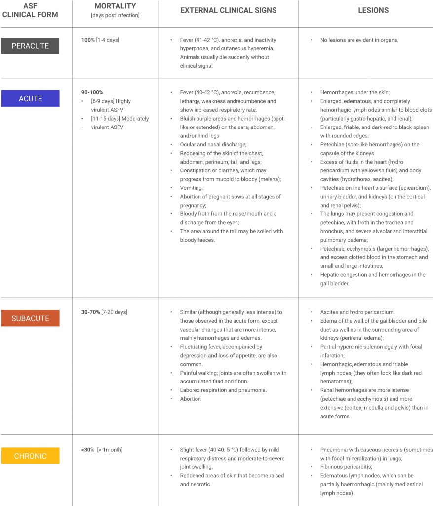
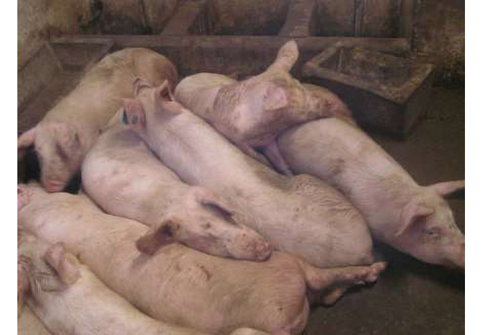
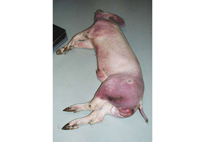
Pathology
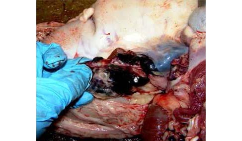
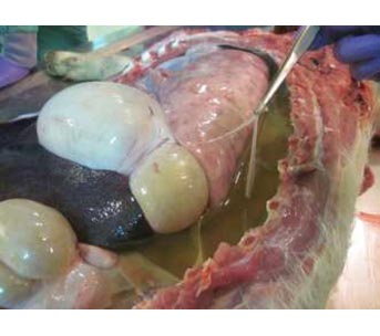
Domestic pigs experimentally infected with African Swine Fever Virus, showing the acute phase of the disease. Typical clinical signs such as anorexia, lethargy and diarrhoea are shown. One pig is painlessly slaughtered, and necropsy is carried out showing typical lesions, like haemorrhagic splenomegaly, haemorrhagic lymphadenitis (mostly in renal and gastrohepatic lymph nodes), petechial haemorrhages in the kidney and urinary bladder together with ascites, hydropericardium, and perirenal oedema.

ENG

![]()
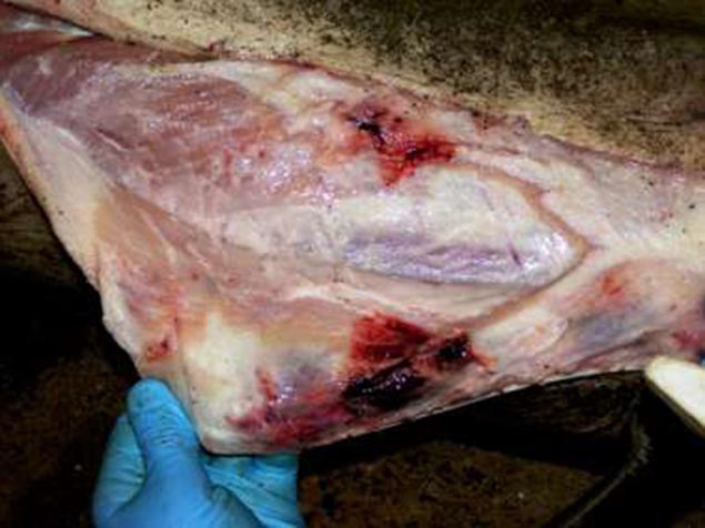
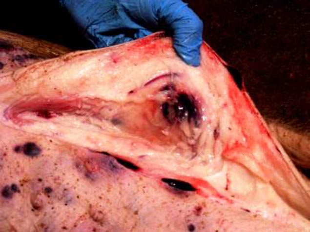
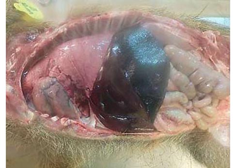
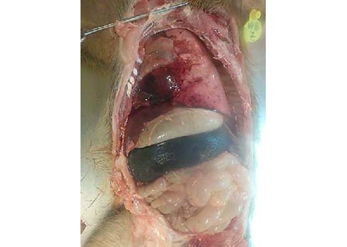
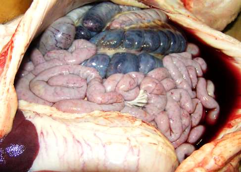
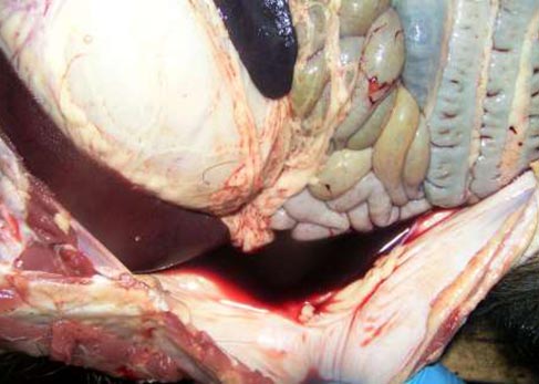
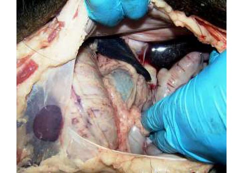
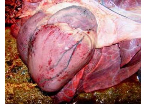
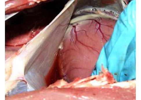
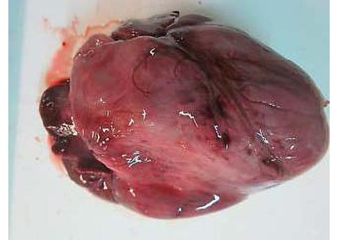
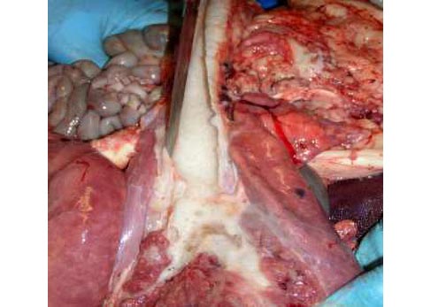
Post-mortem examination of lesions in pigs affected by acute and subacute forms of African swine fever.
EXTENDED VERSION
![]()
![]()
ESP

Muestreo de sangre, fluidos corporales y tejidos de cerdos infectados con el virus de la peste porcina africana para diagnóstico.
![]()
![]()
How does ASF look like?
The present flyer provides essential information about the clinical presentation of the disease to help to identify the most common clinical signs and lesions induced by ASFV Isolates of different virulence.


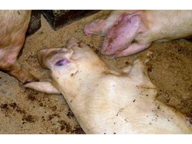
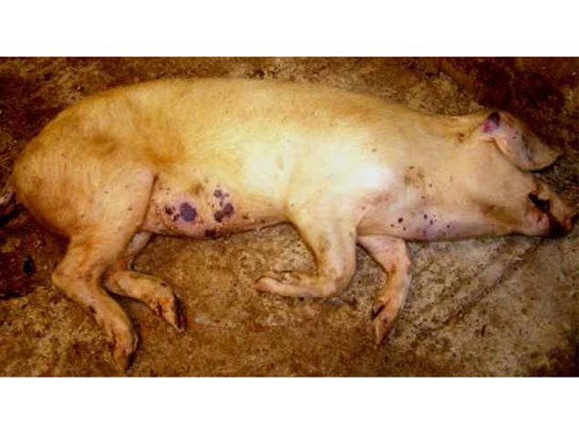
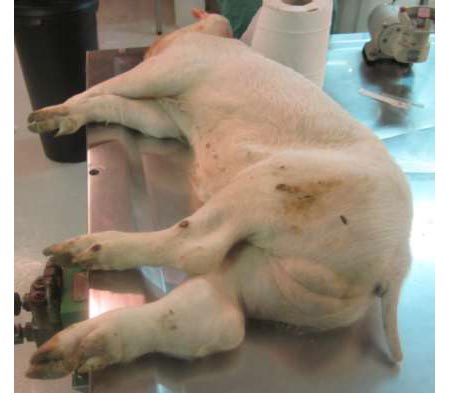
- Pigs are visibly weak with fever and huddle to say warm.
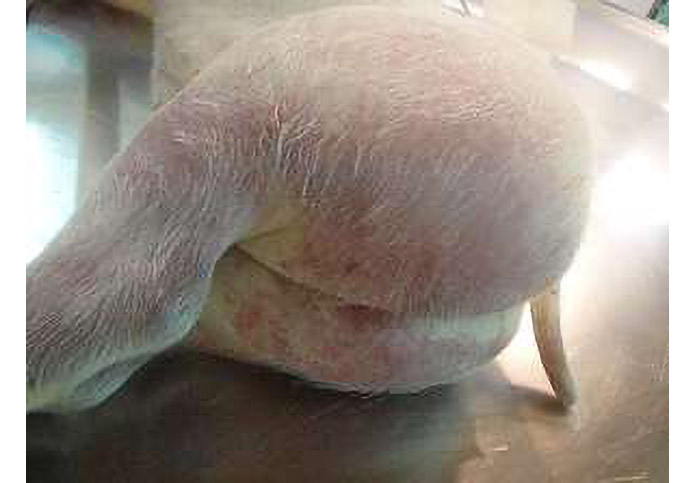
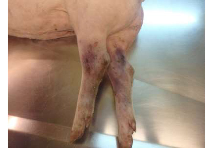
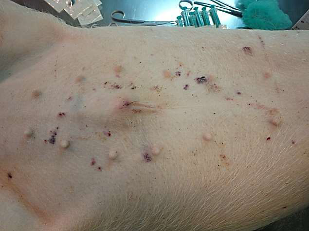
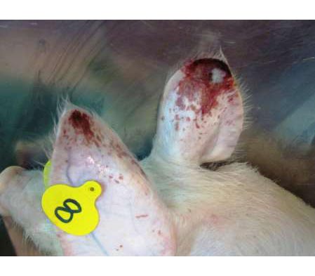
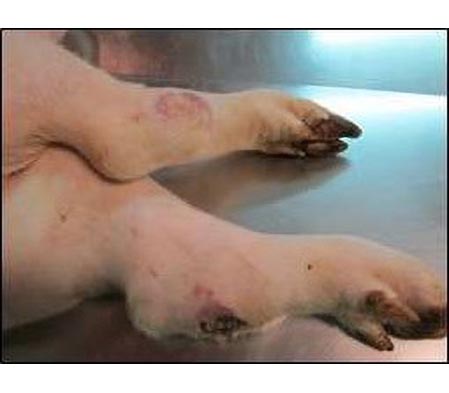
- Reddening of the skin – tips of ears and both front and hind legs.
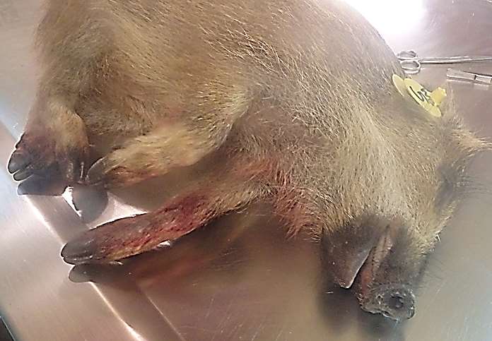
- Cyanosis
- Necrotic areas on the skin surface.
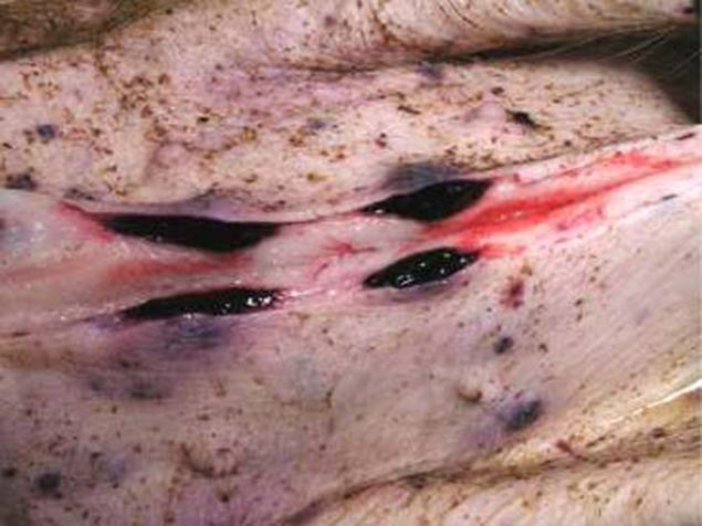
- Subcutaneus haematomas (ears, chest, abdomen and both front and hind legs).
- Melena.
- Epistaxis.
- Foam in mouth/nose.
Abdominal cavity
- Ascites with redish fluid.
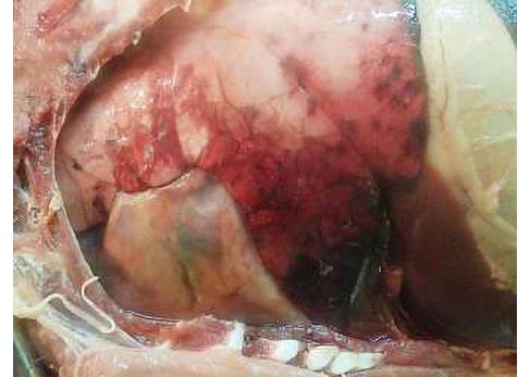
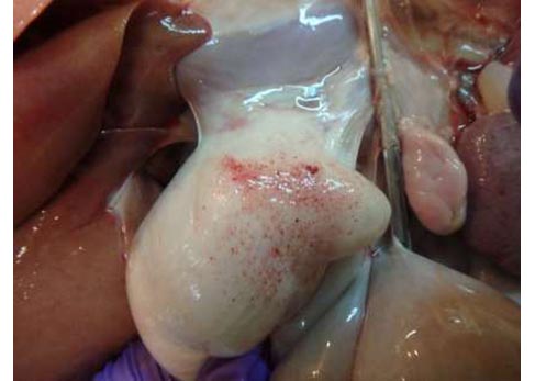
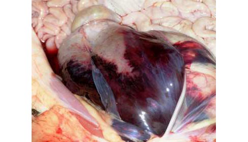
Heart
- Hydropericardium with redish fluid.
- Petechial haemorrhages on epicardium.
- Hydrotorax.
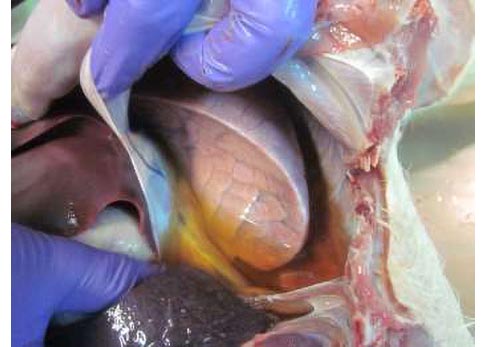
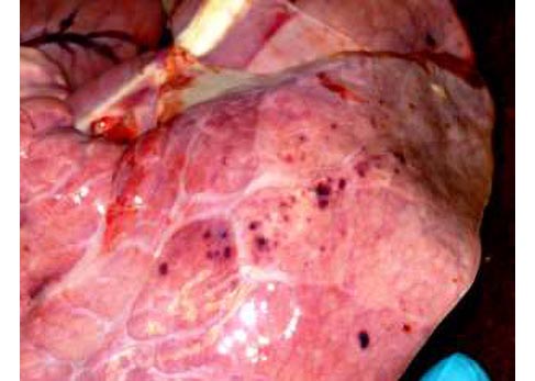
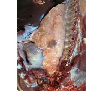
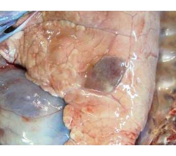
Lung
- Congestión.
- Petechial haemorrhages.
- Froth in trachea and bronchus.
- Severe alveolar and interstitial pulmonary edema.
- Ascites with yellowish fluid.
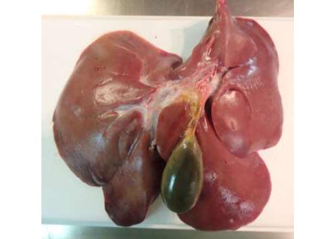
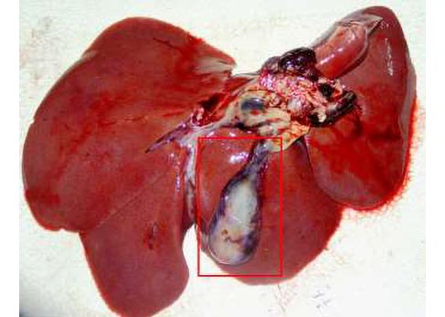
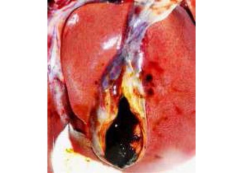
Liver
- Congestión.
- Hepatomegaly.
- Haemorrhages on the serosa Surface of gall bladder.
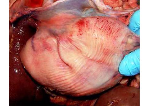
Stomach
- Petechial haemorrhages on serosa and mucosa.
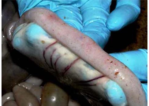
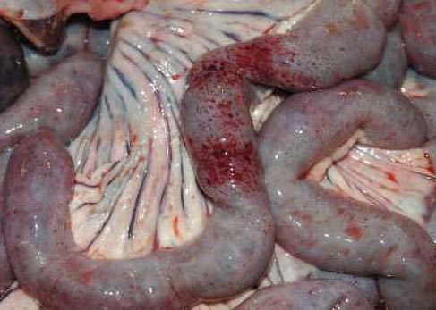
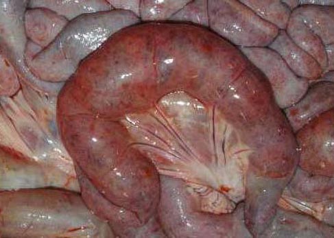
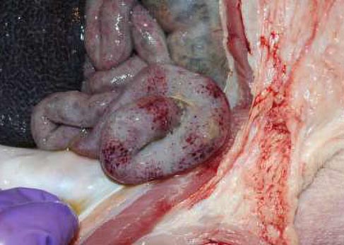
Small and large intestine
- Petechial haemorrhages on serosa and mucosa.
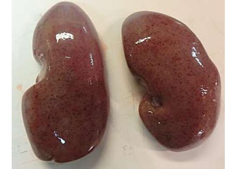
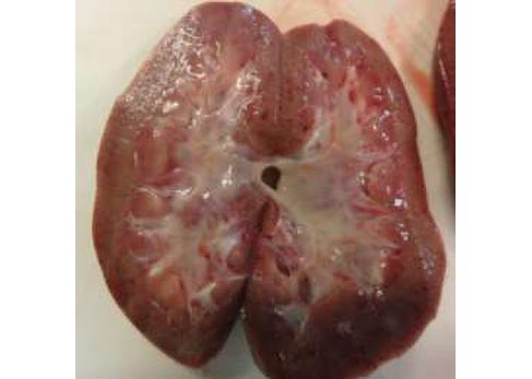
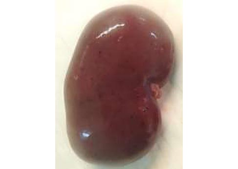
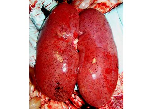
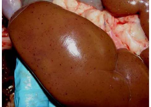
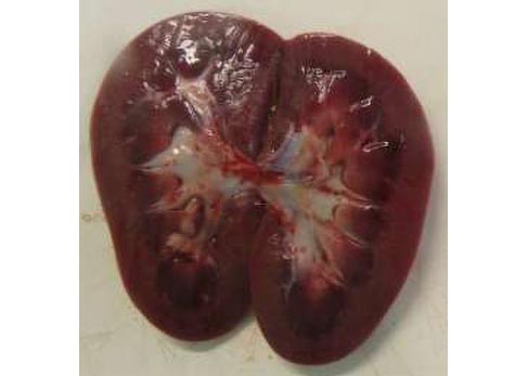
Kidney
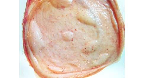
- Petechiae in cortex (more numerous and intense in larger courses of disease).
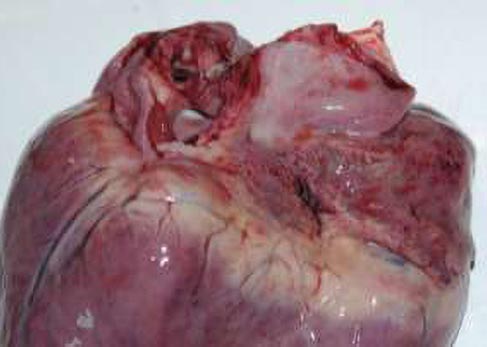
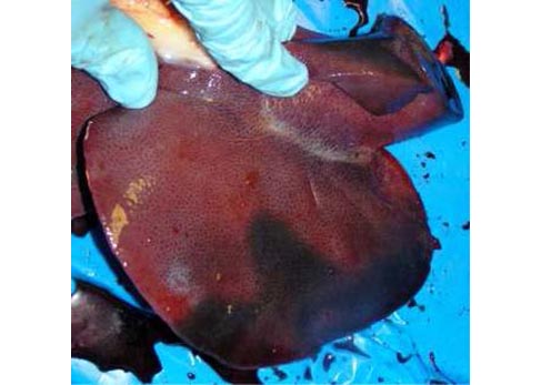
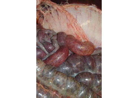
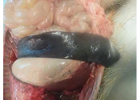
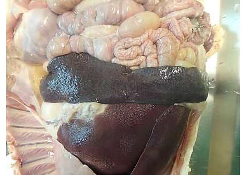
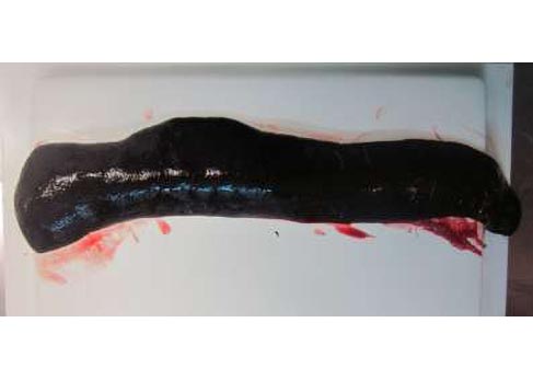
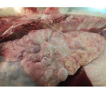
Spleen
- Hyperemic splenomegaly (enlarged with rouded edges, friable and dark red to black).
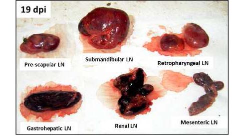
Lymph nodes enlarged edematous and completely hemorrhagic similar to blood clot, mainly gastro hepatic and renal LNs.
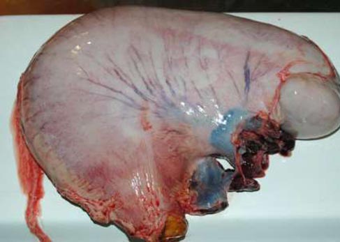
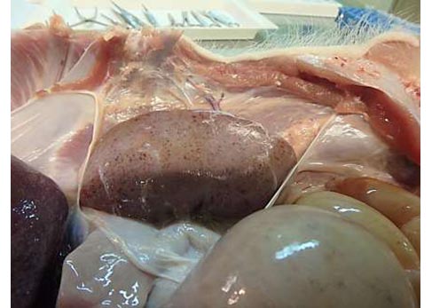
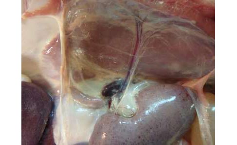
Urinary bladder
- Edema in urinary bladder wall.
- Haemorrhagic (petechiae, ecchymoses and suffusion) in submucosa and serosa.
- Occasional clot blood on mucosa surface.

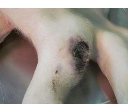
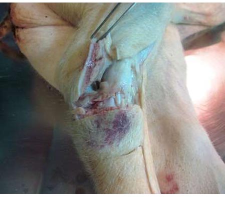
- Moderate to severe joint swelling, often combined with reddened areas of skin that become raised and necrotic.
- Cyanosis.
- Severe respiratory disorders.
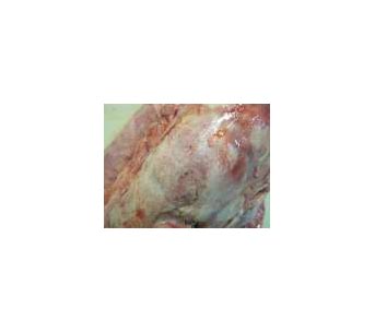
- Caseous necrosis and mineralization of the lungs.
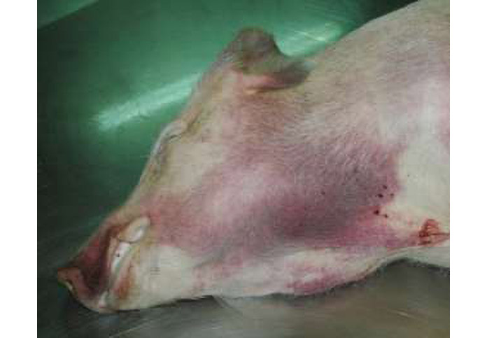
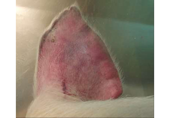
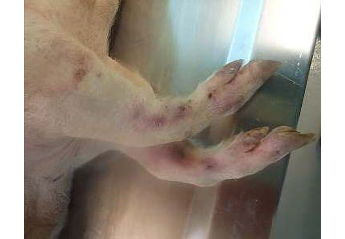
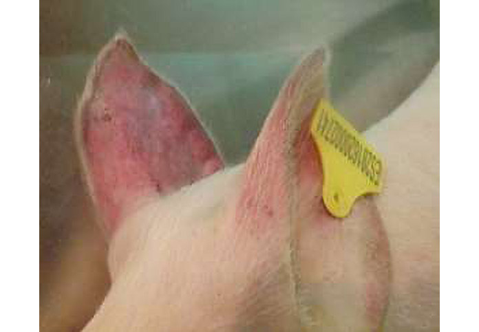
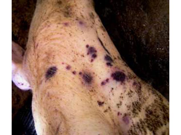
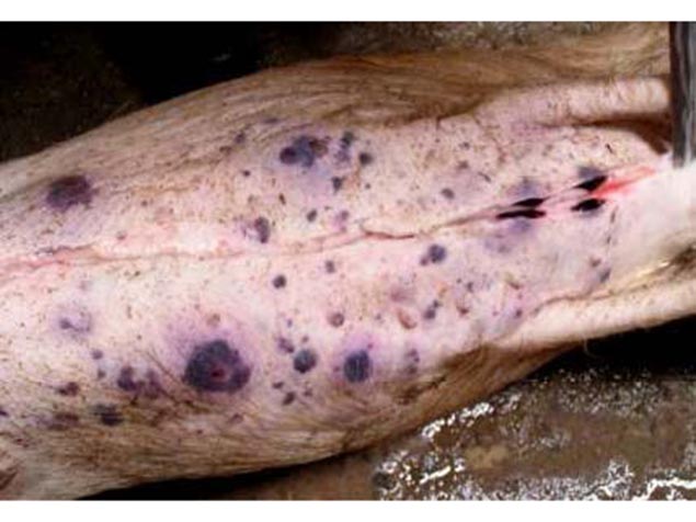
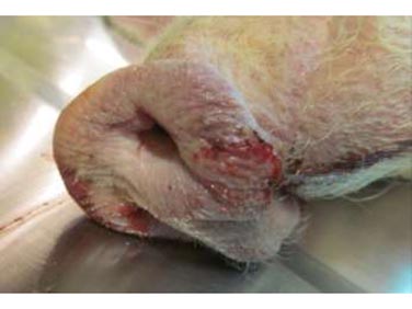
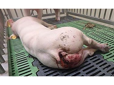
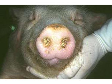
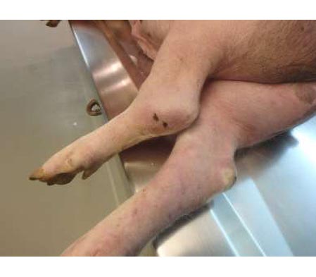
ASF EXTERNAL CLINICAL SIGNS ACUTE-SUBACUTE ASF
- Pigs are visibly weak with fever and huddle to say warm.
- Reddening of the skin – tips of ears and both front and hind legs.
- Cyanosis
- Necrotic areas on the skin surface.
- Subcutaneus haematomas (ears, chest, abdomen and both front and hind legs).
- Melena.
- Epistaxis.
- Foam in mouth/nose.
ASF EXTERNAL CLINICAL SIGNS ACUTE-SUBACUTE ASF
Abdominal cavity
- Ascites with redish fluid.
Heart
- Hydropericardium with redish fluid.
- Petechial haemorrhages on epicardium.
- Hydrotorax.
Lung
- Congestión.
- Petechial haemorrhages.
- Froth in trachea and bronchus.
- Severe alveolar and interstitial pulmonary edema.
- Ascites with yellowish fluid.
Liver
- Congestión.
- Hepatomegaly.
- Haemorrhages on the serosa Surface of gall bladder.
Stomach
- Petechial haemorrhages on serosa and mucosa.
Small and large intestine
- Petechial haemorrhages on serosa and mucosa.
Kidney
- Petechiae in cortex (more numerous and intense in larger courses of disease).
Spleen
- Hyperemic splenomegaly (enlarged with rouded edges, friable and dark red to black).
Lymph nodes enlarged edematous and completely hemorrhagic similar to blood clot, mainly gastro hepatic and renal LNs.
Urinary bladder
- Edema in urinary bladder wall.
- Haemorrhagic (petechiae, ecchymoses and suffusion) in submucosa and serosa.
- Occasional clot blood on mucosa surface.
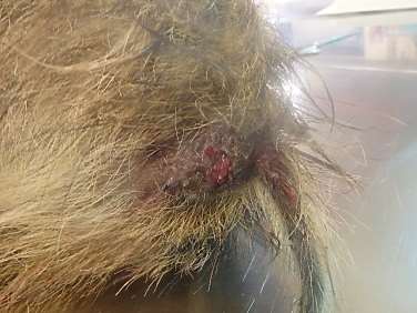
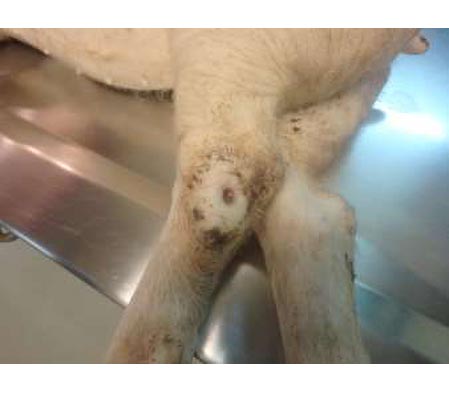
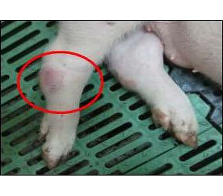
ASF EXTERNAL CLINICAL SIGNS CHRONIC ASF
- Moderate to severe joint swelling, often combined with reddened areas of skin that become raised and necrotic.
- Cyanosis.
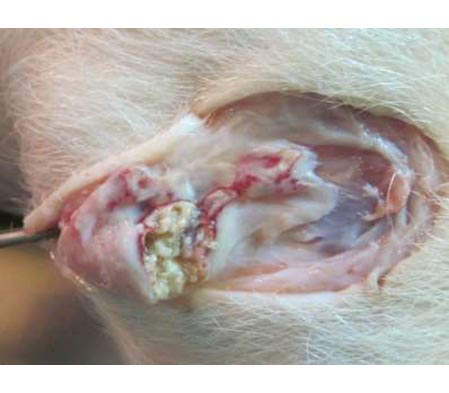
ASF PATHOLOGICAL FINDINGS CHRONIC ASF
- Severe respiratory disorders.
- Caseous necrosis and mineralization of the lungs.